Small Parts Scan: A Detailed Look at Your Body’s Vital Structures
A small parts scan is a specialized ultrasound technique used to evaluate smaller structures and soft tissues in the body. It involves using high-frequency sound waves to capture detailed images of organs, glands, muscles, and other tissues that are not easily visible with conventional imaging methods like X-rays. These scans are particularly useful for diagnosing issues in areas such as the thyroid gland, salivary glands, breasts, testicles, and musculoskeletal system. The detailed imaging provided by a small parts scan helps doctors identify conditions such as lumps, cysts, inflammation, or tumors, which can be crucial for early diagnosis and treatment.
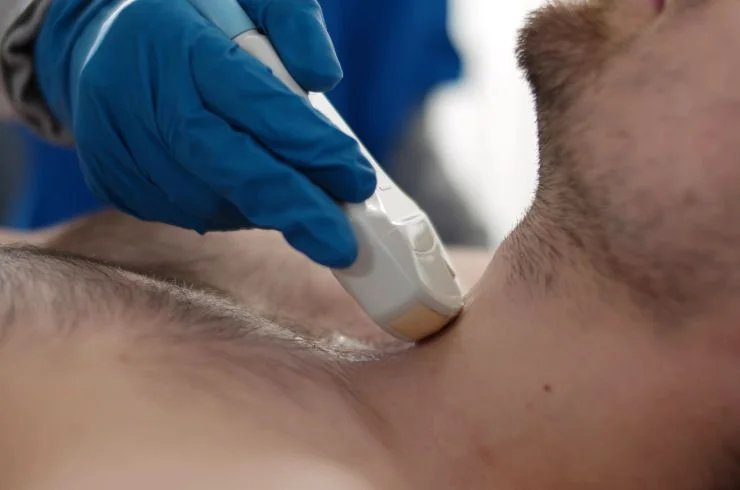
How a Small Parts Ultrasound Works
During a small parts scan, a gel is applied to the skin over the area being examined, ensuring proper transmission of sound waves. A small handheld device called a transducer is then moved over the skin, emitting high-frequency sound waves. These sound waves bounce off the tissues and are converted into real-time images displayed on a monitor. The ultrasound technician or doctor will observe these images to evaluate the condition of the small structures being examined. This non-invasive and painless procedure is quick and offers instant results, providing valuable insights into the health of the area being assessed.
Benefits of Small Parts Scan for Accurate Diagnosis
A small parts ultrasound offers numerous benefits for patients and healthcare providers. It is highly effective in detecting lumps, cysts, and swelling in various parts of the body, making it an essential tool for diagnosing benign or malignant conditions early. For example, it is often used for evaluating breast lumps, thyroid nodules, and testicular masses, helping doctors determine whether further tests or biopsies are necessary. The small parts scan is also non-invasive, avoiding the need for more invasive procedures, and does not require radiation, making it a safer option for repeated imaging. This diagnostic tool provides accurate, real-time images that help doctors make informed decisions, ensuring the best possible care for their patients.
WeCare Diagnostics & Polyclinic, where precision meets care to ensure your health remains our utmost priority. As a leading diagnostic center in Agartala, we offer a comprehensive range of cutting-edge diagnostic services to cater to your medical needs.
Powered By